research using articles via e-resources
Medical
illustrations have been made for thousands of years and mainly used as
documentation, offering accurate records of the body, identifying patients’ condition and diseases, helping medical research, diagnosis and
teaching.
Originally drawings, eventually photography has grown to be an
important aid of medical recording.
On
clinical images, the human body has been used as an object, controlled and
placed into various positions. People, even when their face included, are anonymous,
many illustrations are unisex. In most cases patients can not be identified. This is due to that only
their body parts and innards shown or because of the range of imaging devices and
technologies are used for the captures. This is not always camera but x-ray, ultrasound, magnetic resonance imaging, and other sophisticated machines. The
development of new way of imaging techniques over the years has resulted in the
ability to break down the human body into tissues, bones or even its smallest
cells. Many times the body is not shown as a whole. People are often shown in their most vulnerable state, fighting diseases, opened up during surgery, unconscious or even dead.
Medical
research and teaching need this level of objectivity and representation because
it helps the understanding and the development of science and patient care.
 |
Pituitary tumour as an enlarged
mass in the brain of a 48 year-old female patient
Source: Wellcome Images |
 |
X- ray, Barium meal
Source: Wellcome Images |
The
human body is a fascinating environment and many clinical photographers’ work
cross the line of science and art. While
keeping their educational values, with great artistry and sensitive representation,
they come closer to documentary art.
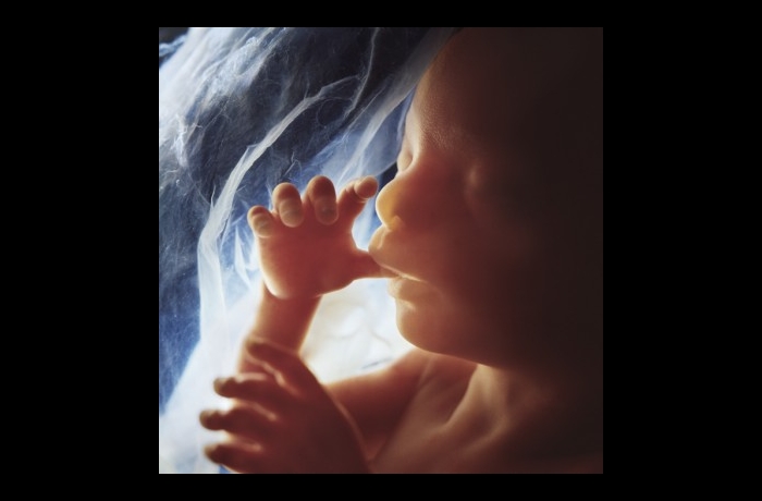
Lennart Nilsson
Lennart Nilsson, pioneering medical
photographer, was born in Sweden, in 1922. Nilsson started out as a
photojournalist during the mid 1940s. During the 1950s, he began to experiment with new photographic
techniques using microscopes. In 1965, his groundbreaking work, The Drama of
Life Before Birth, was the cover story of Life magazine, introducing the public
to unprecedented views of prenatal life, surprising in their clarity and
immediacy. He followed his experiments with photography and light microscopy
with his use of a scanning electron microscope.
Giant Steps 2008, RPS Journal, 148, 4, pp. 150-155, Art Full
Text (H.W. Wilson), EBSCOhost, viewed 21 January 2012
Permalink
His book, A Child is Born, telling
the story of conception to birth. Nilsson's photographs were arguably the most
important images of the 20th century, shedding new light on human life. First
published in 1965, A Child is Born has stayed in print ever since.
Smyth, D 2010, A Child is Born, British Journal Of Photography,
157, 7783, p. 18, Art Full Text (H.W. Wilson), EBSCOhost, viewed 21 January
2012
 |
| Source: www.lennartnilsson.com |
Participating in medical research
and documentation of procedures also turned some non-clinical photographers to
this exciting branch of photography.
Max Aguilera-Hellweg
Max Aguilera-Hellweg spent some twenty years working as a
photojournalist for Roiling Stone, The New York Times and Life. Then, one day
in 1989, he was thrown violently by his horse and had to be operated. While
convalescing, a magazine gave him an assignment, a series of photos of a
surgeon at work. The experience was a revelation. Over the years that followed,
Aguilera-Hellweg photographed some sixty surgical operations. In 1997, the
results were published as The Sacred Heart. The photos are stunning. All were
made following the same formal protocol: the operating theater is dark, with a
beam of light revealing only the part of the body being operated on and the
hands of the surgeon and his team. The powerful light seems to freeze the open
flesh and blood, creating a baroque, Caravaggesque atmosphere. Some of the
images are especially moving, such as the ones showing the separation of two
newborn Siamese twins and the operation on a fetus with spin bifida. Each photo
comes with a text describing the operation for the layman. The most compelling
texts are the artist's own accounts of his experience. Some of his descriptions
are even harder to take than the most brutally raw images, but there is always
a huge respect for humanity.
Leydier, R, & Penwarden, C 2006, Max Aguilera-Hellweg, le
coeur sacré / Opening the Sacred Heart /, Art-Press, 323, pp. 48-49, Art Full
Text (H.W. Wilson), EBSCOhost, viewed 21 January 2012.
 |
| Source: www.maxaugilerahellweg.com |
Theodore Wan
Theodore Wan, medical photographer
and artist’s intriguing series of medical photographs, completed in 1979. The
large format, black and white photographs mimic the technical precision and
visual codes of medical illustration, staging actual diagnostic or preparatory
procedures associated with surgery, with the artist himself positioned as the
patient. In addition to being medical
illustrations these photographs also functioned as art – specifically, by Wan’s
own account, as self-portraiture, and as a form of self investigation. Wan’s
photographs are both art objects, a new kind of self-portraits, and teaching
aids. His body is being constrained and
manipulated into surgical positions, bombarded with x-rays and subjected to the
judgment of others. The way he is feminising his body on some of his images is
curious and raises the question of sexual orientation.
There are also a number of
photographs of Wan alone, and with others, in front of a large grey and white
minimalist grid that functioned a photographic backdrop. As such, it is functioned as a pointed
reference to the Lamprey system of anthropometric photography for purposes of
racial classification, but which also has medical applications.
Conley, C 2008, 'Theodore Wan and the Subject of Medical
Illustration', RACAR: Revue D'art Canadienne/Canadian Art Review, 33, 1/2, pp.
14-27, Art Full Text (H.W. Wilson), EBSCOhost, viewed 21 January 2012.
 |
| Source: cited publication |
Wellcome Centre Medical Image Gallery:
'X-Ray vision' 1997, British Journal Of Photography,
7122, pp. 20-22, Art Full Text (H.W. Wilson), EBSCOhost, viewed 21 January
2012.